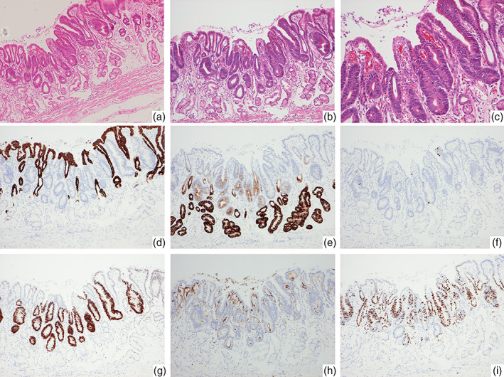
Figure 3.

Histopathological images of Helicobacter pylori‐negative differentiated adenocarcinoma located in the antrum (case 6). (a) The lesion arose from the pyloric glands in the absence of intestinal metaplasia. Fibromuscular obliteration of the lamina propria is observed in the background mucosa. (b,c) The neoplastic gland showing irregular glandular arrangement with low‐grade cellular atypia. There is some nonneoplastic epithelium on the surface layer of the lesion: (b) low power field and (c) high power field. (d–i) Immunohistochemistry: The neoplastic cells showing negative staining for MUC5AC (d) and positive staining for MUC6 (e), MUC2 (f), CDX‐2 (g), and CD10 (h). (i) Ki‐67 labeling index is 59.2%.
