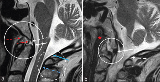
Figure 4.

(a) Sagittal, midline T2 weighted image of the cervical spine demonstrating the anterior atlanto-occipital membrane (red dashed arrow), apical ligament (red arrow), superior longitudinal band of the cruciform ligament (white dashed arrow), tectorial membrane (white arrow), and posterior atlanto-occipital membrane complex, including the posterior atlanto-occipital membrane (blue arrow) and posterior atlanto-axial membrane (blue dashed arrow). (b) Parasagittal T2 weighted image of the cervical spine demonstrating the longus capitis muscle (red star) inserting on the skull base, the alar ligament (white arrow) inserting on the occipital condyle, and the posterior atlanto-occipital membrane (white dashed arrow). Clinical images were obtained from imaging studies performed at our institution. No patient identifying information was included
