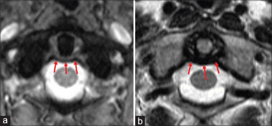
Figure 5.

Axial T2 medic (a) and T2 weighted sequence (b) through C1-C2 level demonstrating the transverse band of the cruciform ligament (red arrows) coursing posterior to the dens and inserting on the inner cortex of the anterolateral C1 arch. The cruciform ligament is the major stabilizing ligament of the atlantoaxial joint. Clinical images were obtained from imaging studies performed at our institution. No patient identifying information was included
