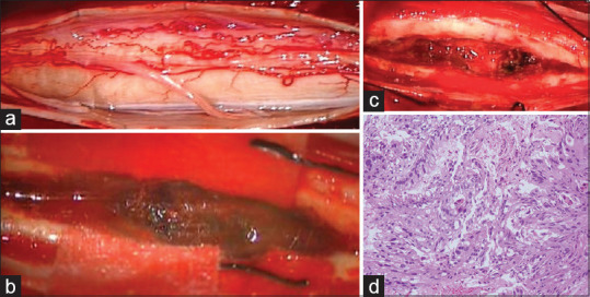
Figure 2.

Intraoperative photographs. Operative exposure showing extensive swelling of the spinal cord itself (a). Myelotomy via a dorsal midline approach showed presence of a grayish-red tumor (b). Gross total resection was successfully performed in a piecemeal fashion (c). Pathological diagnosis after surgery suggested poorly differentiated adenocarcinoma secondary to gastric cancer (H and E, ×400) (d)
