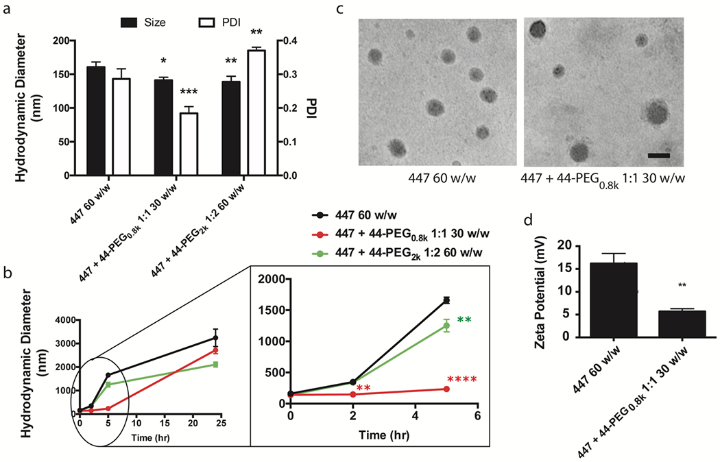
Figure 4. PBAE and PEG-PBAE NP characterization.

(a) Hydrodynamic diameter, polydispersity index (PDI), and (b) 24-h size stability of 447 60 w/w, 447 + 44-PEG0.8k 1:1 30 w/w, and 447 + 44-PEG2k 1:2 60 w/w formulations in artificial cerebrospinal fluid measured by dynamic light scattering (n = 3, mean ± s.d., *: p < 0.05 compared to 447 60 w/w NP). (c) Representative transmission electron microscopy images (scale bar = 100 nm) and (d) zeta potential of 447 60 w/w and 447 + 44-PEG0.8k 1:1 30 w/w formulations (n = 3, mean ± s.d., *: p < 0.05 compared to 447 60 w/w NP).
