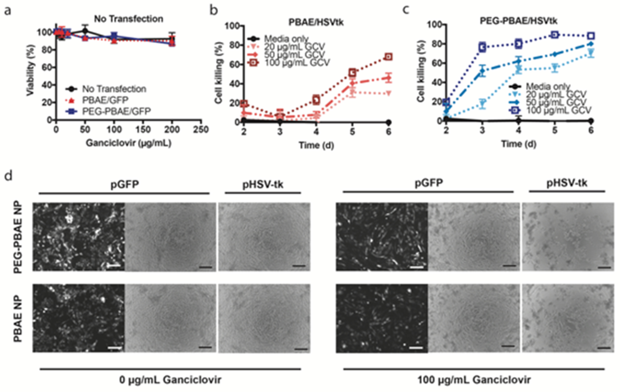
Figure 5. GBM1A cell death mediated by in vitro transfection with HSV-tk and ganciclovir treatment.

Viability of GBM1A cells measured by MTS assay (a) in the absence of HSV-tk transfection after exposure to ganciclovir or (b-c) after HSV-tk transfection using PBAE or PEG-PBAE NPs and incubation with increasing concentrations of ganciclovir (n = 4, mean ± s.d., *: p < 0.05, statistical significance given in Table S2). Cell viability is calculated by normalizing cell counts to those of GFP-transfected cells exposed to the same concentrations of ganciclovir. (d) Representative bright-field microscopy images (scale bar = 200 μm) of GBM1A cells six days after transfection.
