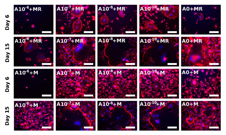
Figure 6.
Visualization of actin ring formation of fused rat peripheral blood mononuclear cells on Days 6 and 15 using epifluorescence microscopy. Filamentous actin was stained with phalloidin (red colour), and nuclei were stained with Hoechst 34580 (blue color). Each image is labeled in the left upper corner by the name of the tested group. The day of visualization is indicated at the beginning of each row. In the presence of both M-CSF and RANKL, the fused rPBMCs were visualized on Day 6 in the A10−7+MR, A10−8+MR, and A10−10+MR groups, indicating the positive effect of ALN in lower concentrations. In the presence of only M-CSF, the fusion of cells began on later experimental days, and, on Day 15, fused cells were observed in A10−7+M, A10−8+M, and A10−10+M groups. Scale bar 100 µm, magnification 200×.

