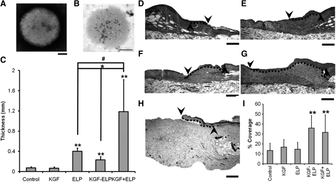
Figure 4.
NPs of fusion protein of ELP and KGF enhance wound healing in diabetic mice by promoting reepithelialization and granulation tissue formation. (A, B) TEM images of KGF-ELP (A) and ELP (B) NPs; scale bar = 100 nm. (C) Quantification of granulation tissue formation in diabetic mice wounds, after the treatment with a fibrin gel, KGF-fibrin gel, ELP-NPs in fibrin gel, ELP-KGF-NPs in fibrin gel, and KGF and ELP-NPs in fibrin gel for 14 days. (D–I) Reepithelialization enhancement of wounds of diabetic mice after treatment. Hematoxylin–eosin staining of wounds after 14 days of treatment with fibrin gel (D), KGF-fibrin gel (E), ELP-NPs in fibrin gel (F), ELP-KGF-NPs in fibrin gel (G), and KGF and ELP-NPs in fibrin gel (H). Dotted line represents the reepithelialization tissue (scale bar of 400 μm). (F) Reepithelialization quantification, normalized to the initial wound gap. Each value represents the mean thickness from 7 mice (n = 7). ** denotes p < 0.05 when compared to control or KGF. # denotes p = 0.043 when compared to ELP particles. * denotes p < 0.01 when KGF-ELP particles are compared with either ELP particles treatment or free KGF + ELP particles treatment. The up arrow indicates the edge of the created wound and the down arrow indicates the tip of the migrating tongue of the wound. Dotted line represents the extent of reepithlialization. Adapted from 84 with permission. ELP, elastin-like protein; KGF, keratinocyte growth factor.

