
FIGURE 3.
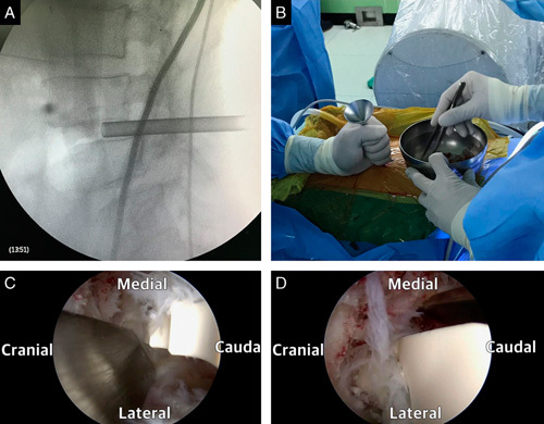
A, Fluoroscopic image of a funnel inserted in the intervertebral disk space. B, Bone grafting through the funnel with the guidance of fluoroscopy. C, Use of semitubular retractor protecting the exiting nerve root during cage insertion. D, Use of semitubular retractor protecting the traversing nerve root during cage insertion.