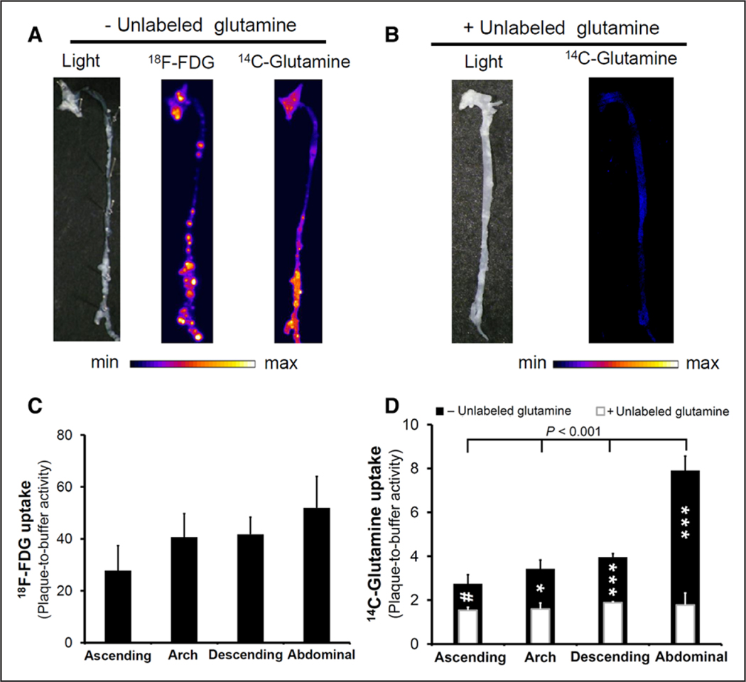
Figure 5.
Autoradiography of atherosclerotic aortas from LDL-R−/− mice fed with a high-fat diet for 3 months. A, Representative images of combined 18F-fluorodeoxyglucose (18F-FDG) and 14C-glutamine ex vivo autoradiography demonstrated different patterns of substrate accumulation in aortic atherosclerotic plaques. B, The specificity of 14C-glutamine uptake was determined through the coincubation with 20-mmol/L unlabeled glutamine, which resulted in a noticeable decrease in uptake throughout the aorta. C, The maximal 18F-FDG plaque accumulation was not statistically different between various segments of the aorta. On the contrary, 14C-glutamine accumulation (D, black bars) was significantly higher in the abdominal aorta compared with the remaining segments. Consistent with the visual assessment of plaque accumulation, coincubation with unlabeled glutamine (D, overlaid white bars) resulted in a statistically significant decline in quantitative maximal 14C-glutamine uptake throughout the aorta. (n=6 for nonblocking and 3 for blocking experiments). (#P=0.07, *P<0.05, and ***P<0.001 maximal plaque accumulation in the presence vs absence of unlabeled glutamine).

