Fig. 1.
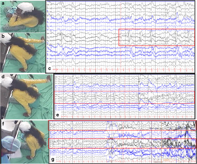
(a) Child is sitting normally, while in (b), her right arm is seen going backward in a dystonic manner. The corresponding EEG (c) shows rhythmic spike and slow waves seen at a higher frequency, along with some lower amplitude rhythmic activity in the theta range. (d) The child has sustained posturing of the right arm in the ongoing seizure. (e) The corresponding EEG shows well-evolved rhythmic slow waves in the left frontal and temporal chains. (f) The seizure has ended here, obvious by the right arm of the child now by her side and recovered from the dystonic posturing. (g) In the first half of the panel, the left-sided channels continue to have rhythmic delta activity, which become higher in amplitude and even slower as the seizure ends, in the second half of the epoch shown

