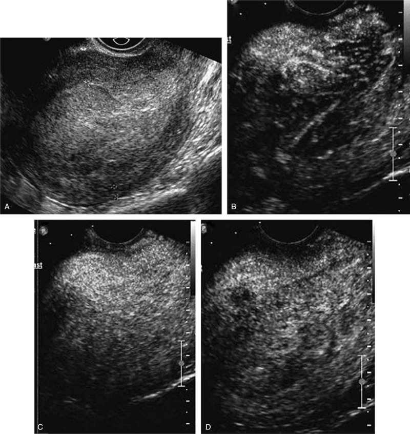
Figure 9.

Images of a stage IA endometrial carcinoma with bulky tumor. (A) Image before the injection of the bolus showed marked enlargement of endometrium. (B) Image at 14 seconds after administration showed asymmetrically enhanced tumor. (C) Image at 18 seconds showed the maximal concentration of contrast agent in the tumor. (D) Image at 27 seconds showed that tumor washed out earlier than normal myometrium, thus formed a clear peritumoral halo.
