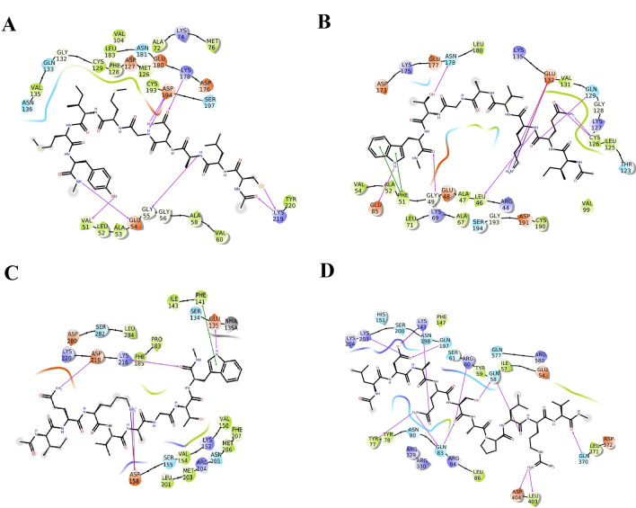
Fig. 5.
Interactions established after docking, a CLANGMIMY-AAK1, b IQKVAGTW-GAK, c IQKVAGTW-PIKfyve and d LDAQSAPLRV-TPC2. Peptides are in green, while residues of binding sites are represented in three coded format (various color) and thick colored lines encasing peptides represent the active protein site pockets. Solid pink lines depict H-bonds, while hydrophobic interactions are represented in green. Additionally, salt bridges and π-cation stacking, are represented by gradient color (pink and blue lines) and a red line respectively (Color figure online)

