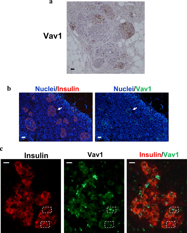
Fig. 2.
In normal human pancreatic tissue Vav1 is present in insulin producing cells. Representative images of formalin-fixed paraffin embedded normal pancreatic tissue sections subjected to immunohistochemical analysis with anti-Vav1 antibody (A) and to immunofluorescence using simultaneously antibodies against Vav1 (green staining) and insulin (red staining). Nuclei were counterstained with DAPI (B, C). In B, overlay of Vav1 (green) or insulin (red) with nuclei (blue) is reported. The arrow indicates a pancreatic islet positive for both Vav1 and insulin. In C, merged insulin/Vav1 staining is shown with co-localization resulting in yellow. The dashed rectangles identify β-cells with various levels of insulin and Vav1. Bar = 50 μm

