Fig. 5.
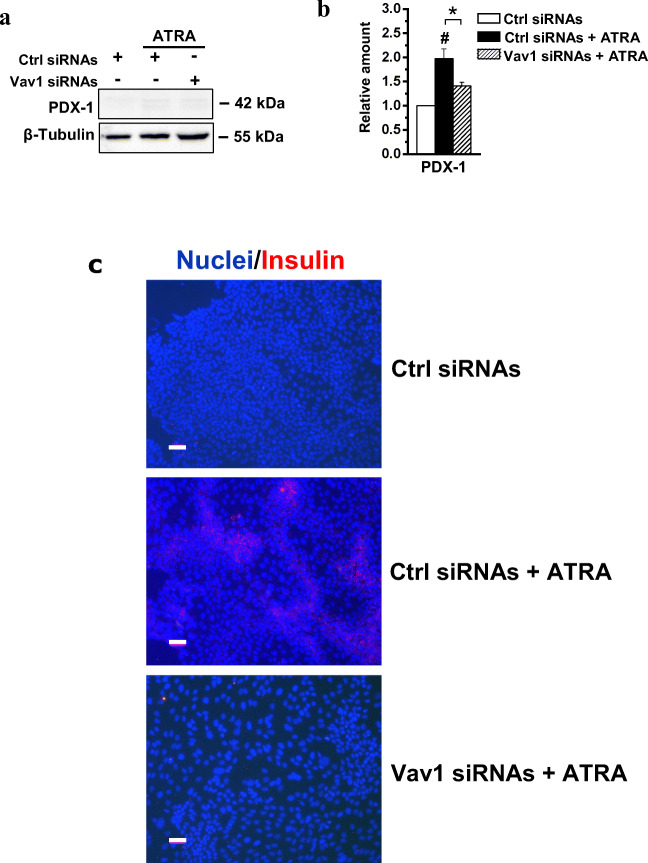
Vav1 sustains the ATRA-induced trans-differentiation of HPAF-2 cells. (A) Representative immunochemical analysis performed with the indicated antibodies of lysates from HPAF-2 cells transfected with siRNAs specific for Vav1 (Vav1 siRNAs) and cultured for 96 h in the presence of 1 μM ATRA. Scramble siRNAs (Ctrl siRNAs) was used as control. (B) Levels of PDX-1 as deduced from the densitometry of immunochemical bands normalized with β-Tubulin, used as internal control for equivalence of loaded proteins. All the data are the mean of three separate experiments performed in triplicate ±SD. # P < 0.05 versus control; * P < 0.05 between bars. (C) Representative immunofluorescence staining of HPAF-2 cells grown on glass dishes for 96 h under the same experimental conditions and subjected to immunocytochemical analysis with the anti-insulin antibody (red staining). Nuclei were counterstained with DAPI (blue staining). In all the examined experimental conditions, merged images of insulin expression with nuclear staining (Nuclei/Insulin) are shown. Bar = 50 μm

