Figure 6.
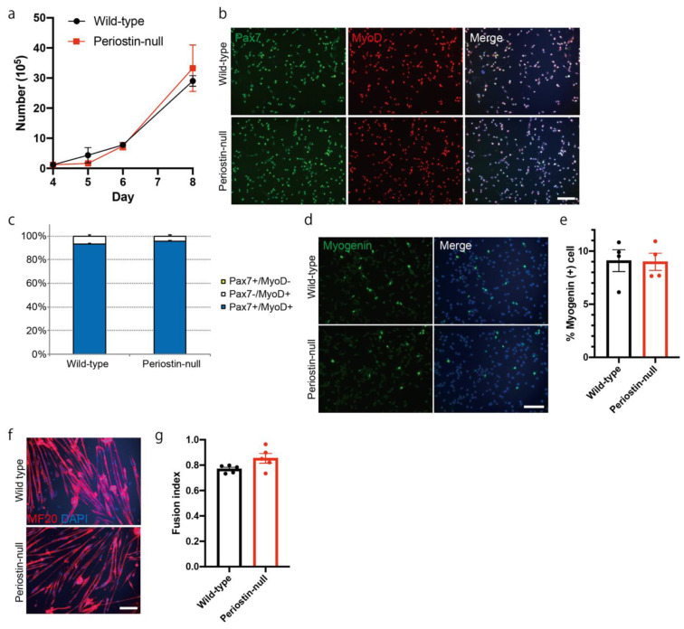
Proliferation and differentiation of muscle progenitor cells are not impaired in periostin-null mice. (a) Time-course changes in the number of cultured muscle progenitor cells. n = 4. (b) Immunocytochemical analysis of Pax7 and MyoD in muscle progenitor cells from periostin-null mice. Bar: 200 μm. (c) Ratio of Pax7+/MyoD-, Pax7-/MyoD+, and Pax7+/MyoD+ cells. n = 4. (d) Immunocytochemical analysis of myogenin in muscle progenitor cells from periostin-null mice. Bar: 200 μm. (e) Quantitative analysis of myogenin-positive cells. n = 4. (f) Immunocytochemical analysis of MF-20 in differentiated muscle progenitor cells from periostin-null mice. Bar: 200 μm. (g) Quantitative analysis for the fusion index. n = 5. Error bars indicate SEM.

