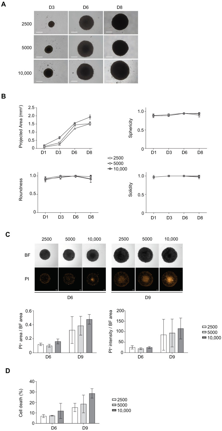
Figure 1.
Influence of cell density on ultra-low attachment (ULA)-multicellular aggregates of lymphoma cells (MALC) biology. ULA-MALC established with RL cells was cultured at different cell seeding densities (2500; 5000 and 10,000 cells) and different biological characteristics were determined at different culture times. (A) Growth and morphology observed by bright field (BF) microscopy at 4× after 3, 6 and 8 days (D) of culture. Scale: 500 µm. These pictures are representative of 3 independent experiments each comprising 6 individual ULA-MALC. (B) Morphological properties (projected area, sphericity, roundness and solidity) determined after 1, 3, 6 and 8 days (D) of culture by 2D imaging analysis with the specific macro developed (see Material and Methods section). These graphs are the mean ± SD of n = 3 independent experiments each comprising at least n = 10 individual ULA-MALC. (C) Cell death visualization and quantification on whole ULA-MALC at day 6 and 9 following propidium iodide (PI) labeling and 2D imaging. Experiment performed on 3 independent experiments each comprising 7 individual ULA-MALC. Upper panel, representative pictures of bright field (BF) or propidium iodide (PI) at 5× magnification, scale: 200 µm. Lower panel, mean ± SD of the ratio of area or intensity of propidium iodide (PI) in relation to the bright field (BF) area. (D) Cell death quantification by flow cytometry after 7AAD labeling of dissociated MALC. This graph represents mean ± SD of the percentage of cell death (7AAD+) measured in 3 independent experiments of 3 pooled ULA-MALC.

