Figure 5.
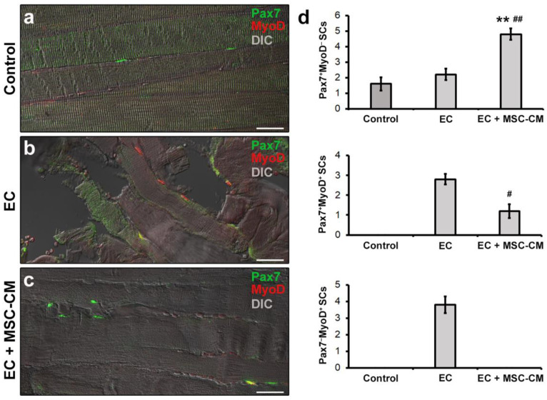
Morphological evaluation of satellite cells (SCs) in formalin-fixed and paraffin-embedded tissue sections from control EDL muscles, EDL muscles damaged by ex vivo forced eccentric contraction (EC) and EDL muscles damaged by EC in the presence of bone marrow-mesenchymal stromal cell (MSC) conditioned medium (EC + MSC-CM). (a–c) Representative superimposed confocal immunofluorescence and differential interference contrast (DIC, grey) images of muscle tissue sections immunostained with antibodies against the SC marker Pax7 (green) and the SC activation marker MyoD (red). Quiescent SCs or SCs likely contributing to replenish SC basal pool are identifiable as Pax7+/MyoD− (green), tissue damage-activated SCs as Pax7+/MyoD+ (yellow), and SCs committed into the myogenic program as Pax7−/MyoD+ (red). Images are representative of at least 3 sections from each of 4 control, 4 EC and 4 EC + MSC-CM muscles. Scale bars = 25 μm. (d) Quantitative analysis of Pax7+/MyoD−, Pax7+/MyoD+ and Pax7−/MyoD+ SCs in the different experimental conditions. Values are mean ± SEM. ** p < 0.01 vs. control; # p < 0.05 and ## p < 0.01 vs. EC.

