Figure 2.
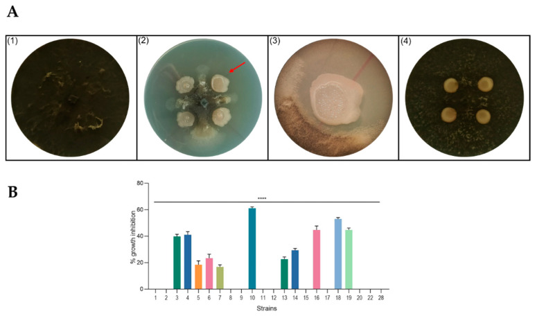
Antagonism assays in solid medium. (A) Representative photographs of dual-culture assay for in vitro inhibition of mycelial growth of M. phaseolina by isolated strains. (1) M. phaseolina (control plate); (2) example of active strain (RHFS10) against M. phaseolina growth; (3) images of interaction zone of RHFS10 strain and M. phaseolina acquired with a stereoscopic microscope (10× magnification); (4) example of inactive strain (RHFS28) against M. phaseolina growth; red arrow in panel 2 indicates the interaction zone magnified in panel 3. (B) Inhibition of fungal growth reported as the percentage reduction in the diameter of the fungal mycelia in the treated plate compared to that in the control plate. All experiments were performed in triplicate with three independent trials. Data are presented as means ± standard deviation (n = 4) compared to control M. phaseolina grown without bacteria. For comparative analysis of groups of data, one-way ANOVA was used and p values are presented in the figure: ****: extremely significant < 0.0001.

