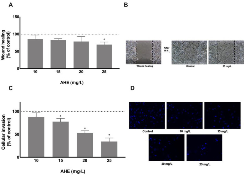
Figure 3.
The ability to impair HMEC-1 migration and invasion was performed after 16 h and 24 h treatment with AHE by injury assay (A,B) and matrigel-coated transwell assay (C,D), respectively. There is an abrogation in cell motility and the capacity to migrate to the wound area was reduced at a concentration of 25 mg/L (A). Dotted lines represent the wound healing area at the beginning of experiment (B). HMEC-1 invasive capacity through a matrigel membrane was reduced in a dose-dependent manner with significance for doses of 15 mg/L or higher (C). Representative images of invasive HMEC-1 under fluorescence microscope (D). Results are presented as % of control group mean ± SD. * p < 0.05 vs. control.

