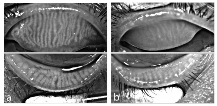
Figure 2.
Infrared meibography prior to aHSCT (a) OS of female patient, age 58, acute myeloid leukemia with normal distribution of meibomian glands, (b) OD of male patient, age 52, chronic myeloid leukemia with extensive loss of meibomian glands in both eyelids (Meibographer: Keratograph 5M, OCULUS Optikgeraete GmbH, Wetzlar, Germany).

