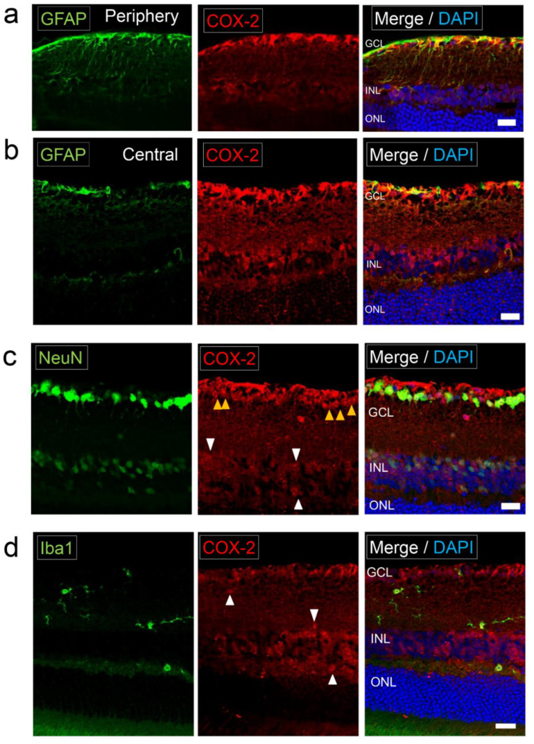
Figure 2.
Expression of COX-2 in normal retina. Photomicrographs of the retina in normal C57BL/6J mice (a) The retinal section of the peripheral area labeled with GFAP (astrocyte/Müller cell marker) and COX-2. COX-2 was positive in GFAP+ cells. GFAP-positive vertical lines (Müller cells) were not clearly COX-2 positive. Scale bar, 20 μm. (b) The retinal section of the central area labeled with GFAP and COX-2. COX-2 staining coincided with GFAP staining. Scale bar, 20 μm. (c) The retinal section labeled with NeuN (neuronal marker) and COX-2. COX-2 was positive in NeuN+ neurons in the GCL (yellow arrowheads) and the INL (white arrowheads). Scale bar, 20 μm. (d) The retinal section labeled with Iba1 and COX-2. Iba1+ cells were ramified microglia with processes. COX-2 was weak positive in Iba1+ ramified microglia (white arrowheads). Scale bar, 20 μm.

