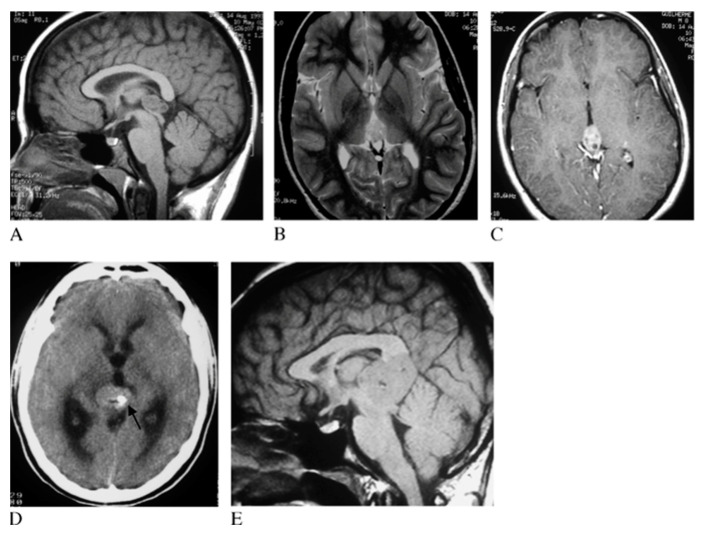
Figure 3.
Pineal germinoma magnetic resonance imaging (MRI). Pineal tumor hypointense on T1 weighted image (T1WI) (A), hyperintense on T2WI (B) and with homogeneous contrast enhancement (C). The arrow identifies hyperdense mass with calcification at computed tomography (D). In another patient, a larger germinoma, isointense on T1WI (E). Reprinted with permission from Reis et al. (2006) [41]. John Wiley and Sons—2021 (License Number 5034711197310).

