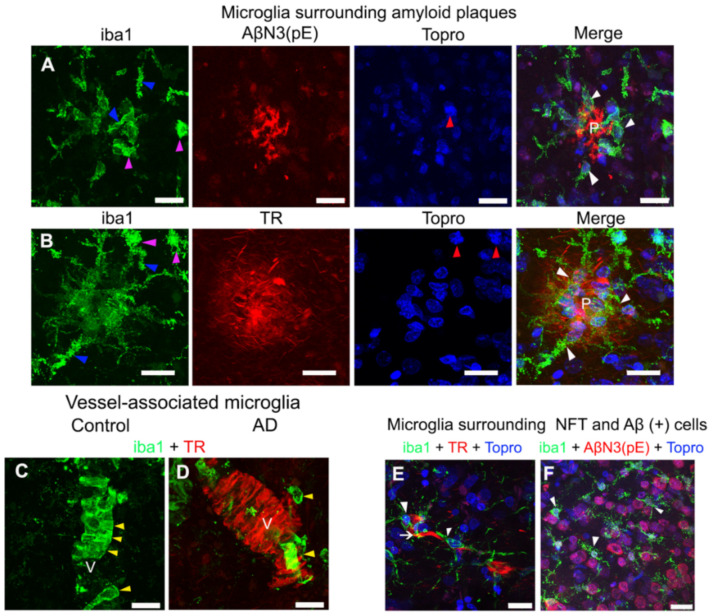
Figure 2.
Representative micrographs of double and triple immunostainings of microglia in AD brains. (A) Microglia (green channel) surrounding (white arrowheads) an amyloid plaque (P) with AβN3(pE) peptide deposits (red channel). (B) Microglia (green channel) surrounding (white arrowheads) a fibrillar TR-positive amyloid plaque (P; red channel). Changes in the morphology (purple arrowheads) of microglia ramifications (blue arrowheads) and the nuclear morphology (blue channel; red arrowheads) are indicated in both panels. Control (C) and AD (D) brains show vessel (V)-associated microglia (yellow arrowheads; green channel) counterstained with TR (red channel). (E,F) Representative micrographs of microglia (white arrowheads; green channels) surrounding a neurofibrillary tangle (NFT) (white arrow; red channel) and Aβ (+) cells (red channel), respectively. Some tissues were counterstained with TO-PRO®-3 (blue channel). The scale bar = 20 μm.

