Figure 5.
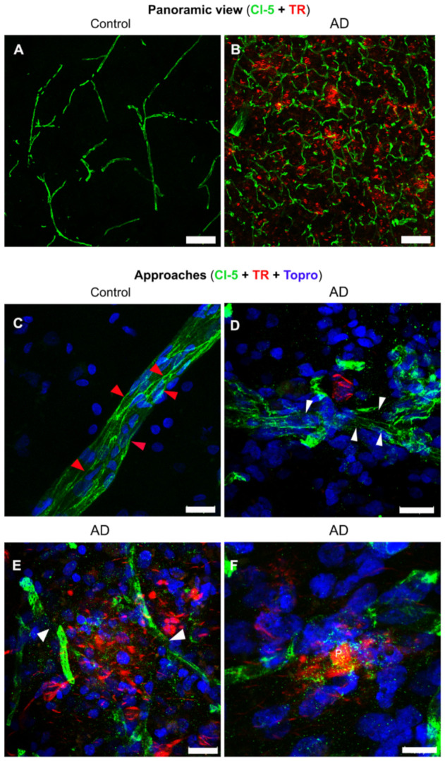
Double and triple immunofluorescent staining of tight junctions in AD and control brains. Representative panoramic views of control (A) and AD (B) brains with double immunostaining for claudin- 5 (Cl-5; green channels) and TR (red channels). (C–F) Representative images of triple immunostaining in control (C) and AD (D–F) brains, for Cl-5 (green channels), TR (red channels), and TO-PRO®-3 (blue channels). Continuity (red arrowheads) and discontinuity (white arrowheads) of the tight junctions are indicated. The scale bars = 100 µm for the panoramic views (A,B) and 20 μm for magnifications (C–F).

