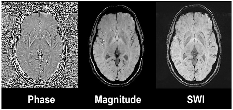
Figure 1.
Susceptibility weighted image (left) is the result of the combination of high-pass filtered phase (left) and magnitude image (central). Axial slices at the level of basal ganglia on a 3 Tesla Siemens Magnetom Trio system with 32-channel phased-array head coil. Reprinted with the permission of [15].

