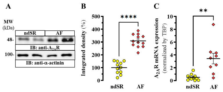
Figure 1.
A2AR expression in human right atrium. (A) Representative immunoblot showing the expression of A2AR in right atrium from non-dilated sinus rhythm (ndSR) and atrial fibrillation (AF) patients; membranes from human atrium were analysed by SDS-PAGE (10 μg of protein/lane) and immunoblotted using goat anti-A2AR and rabbit anti-α-actinin antibodies (see Methods). (B) Relative quantification of A2AR density; the immunoblot protein bands corresponding to A2AR and α-actinin from ndSR (n = 11) and AF (n = 12) patients were quantified by densitometric scanning; values were normalized to the respective amount of α-actinin in each lane to correct for protein loading. (C) Relative expression of A2AR transcripts in human atrium. Mean ± SEM from ndSR (n = 11) and AF (n = 9) patients. **** p < 0.0001 and ** p < 0.01, Student t test.

