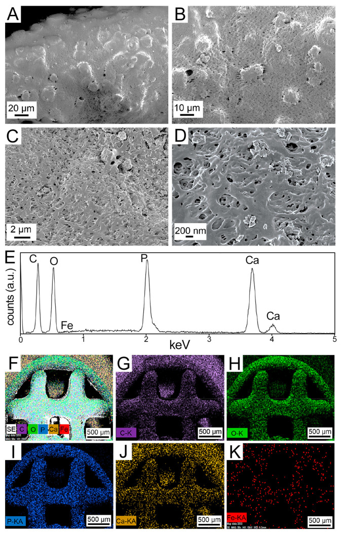
Figure 2.
Material characterization analyses conducted on bioprinted 60 µg/mL SPION-loaded HB scaffolds. (A–D) Field-emission scanning electron microscopy (FESEM) images of the SPION-loaded 3D bioprinted HB scaffold at varying magnifications. (E–K) Energy-dispersive X-ray spectroscopy (EDX) spectrum of the HB-SPION scaffold (E), along with the EDX maps of (F) secondary electrons, (G) carbon (C), (H) oxygen (O), (I) phosphorous (P), (J) calcium (Ca), and (K) iron (Fe) in the printed constructs.

