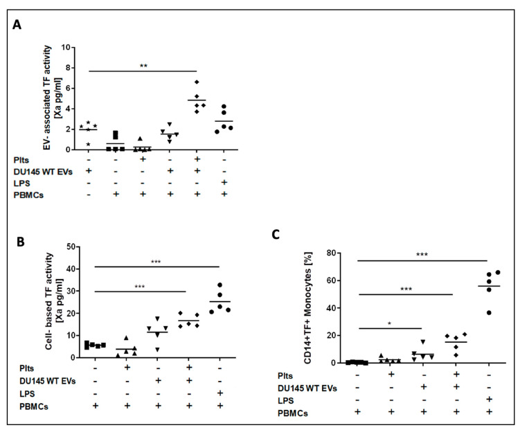
Figure 4.
Co-incubation of conditioned media from TF-exposing DU145 prostate cancer cells with peripheral blood mononuclear cells (PBMCs) and platelets. EV-TF activity was detectable in conditioned media from the DU145 cell line. Low EV-TF activity was found in conditioned media from PBMCs alone and PBMCs that were co-incubated with platelets. Co-incubation of PBMCs with DU145 EVs did not significantly increase EV-TF activity. EV-TF activity significantly increased when DU145 EVs, PBMCs, and platelets were co-incubated (A). Cell-based TF activity was detectable but low on unstimulated PBMCs and PBMCs that were co-incubated with platelets. Cell-based TF activity significantly increased by co-incubation of PBMCs with EVs from DU145 cells and further increased when platelets were added (B). By applying flow cytometry, we found that only monocytes (CD 14+) expose TF but not lymphocytes (CD14−) after co-incubation with DU145-derived EVs and platelets (C). Cell-based TF activity (pg/mL) was determined in triplicate. Each dot represents a single measurement and the line represents the mean. (* p ≤ 0.05, ** p ≤ 0.01, *** p ≤ 0.001, paired comparison for repeated measurements). Abbreviations: LPS—lipopolysaccharide.

