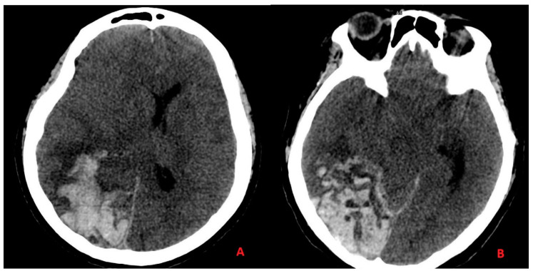
Figure 2.
Acute ischemia with hemorrhagic infarction in the absence of any vascular malformation with acute quote of cerebral edema showed on CT head scan images. In panel (A), we can see the hemorrhagic infarction of the parietal and temporal lobes, with edema on the white matter of the antero-lateral portion of the temporal lobe with added “mass effect” on the adjacent structures. In panel (B), we can see the hemorrhagic infarction of the occipital and temporal lobes, and the subarachnoid hemorrhage on the cerebellar tentorium margin.

