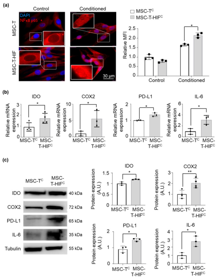
Figure 3.
HIF overexpression strengthens signaling through the NF-κB pathway and consequently the expression of immunosuppressive cytokines. (a) Immunolocalization of p65 in MSC-T, MSC-Tc, MSC-T with an HIF-1α-GFP lentiviral vector (MSC-T-HIF) and conditioned MSC-T-HIF (MSC-T-HIFc). Red: p65, blue: DAPI. Scale bar: 30 μm. Mean of fluorescence intensity was measured per nucleus and values were normalized to those of MSC-T cells (each point represents the mean of 50 nuclei). Graphs represent mean ± SD of three independent experiments. Sidak’s multiple comparisons test was used for statistics. (b) IL-6, IDO, COX2 and PD-L1 expression levels quantified by RT-qPCR in MSC-Tc and MSC-T-HIFc. Expression level of the target gene in each sample was normalized to GAPDH expression. Graphs represent mean ± SD of fold change of three independent experiments. Paired t-test was used for statistics. (c) Representative Western blots of IL-6, IDO, COX2 and PD-L1 proteins. Expression levels were quantified by densitometry relative to MSC-Tc. α-tubulin was used as a protein loading control. Bars represent mean ± SD of three independent experiments. Paired t-test was used for statistics. * p < 0.05, p < 0.01.

