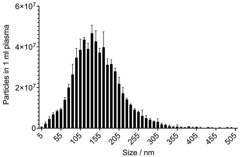
Figure 2.
Biological triplicate of extracellular vesicles (EVs) from three cancer patients isolated with the EXÖBead® technique, showing the typical size range of exosomes between 30–150 nm. Average particle concentration with standard deviation in 1 mL plasma: 6.11 × 10⁸ ± 4.41 × 10⁷. Average particle size with standard deviation in 1 mL plasma: 138.30 ± 2.59. Analysis with a ZetaView® (Mebane, NC, USA): Nanoparticle Tracking Analyzer.

