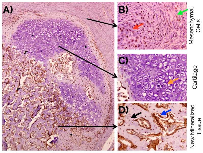
Figure 6.
(A) Overview of RUNX2 detection in a bone fracture callus (in brown) and detail observations in (B) fusiform mesenchymal cells (green arrow), and immature chondrocytes (red arrow) in the fibrocartilage, (C) negative detection in mature hypertrophic chondrocytes (orange arrow), and (D) intense detection in the nucleus and cytoplasm of osteoblasts (blue arrow), and osteocytes (black arrow) in the newly formed bone. Peroxidase-conjugated micropolymer detection in samples from the 14-day time point. Original magnification: 10× (A) and 20× (B–D).

