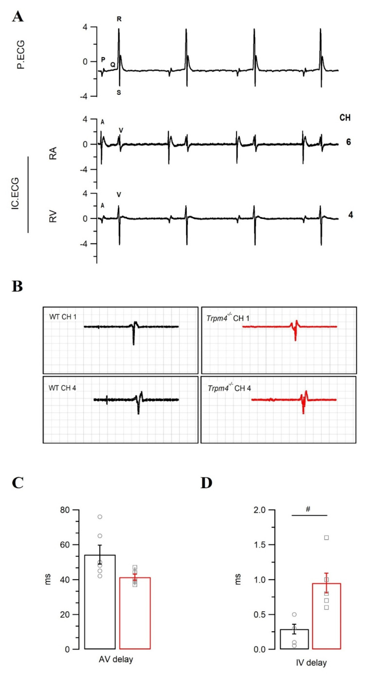
Figure 5.
Intracardiac electrocardiogram recordings on WT and Trpm4−/− explanted hearts. (A) Representative pseudo (P.ECG) and intracardiac (IC.ECG) ECG trace from a WT heart. A and V respectively represent atrial and ventricle signals from the intracardiac catheter recorded in different electrode channels (CH 1-7). Conduction delays either between the atria and ventricle (AV delay, CH 6-5) or intraventricular (IV delay, CH 4-1) were measured as the time delay between respective channel signals. Representative trace showing signal derivatives from CH 1 and 4 to measure IV delay (B). Average AV delay (C) and IV delay (D), compared between the genotypes (WT: circle, black and Trpm4−/−: square, red). Note: N = 6, #: p < 0.05, WT vs. Trpm4−/−; ns = not significant.

