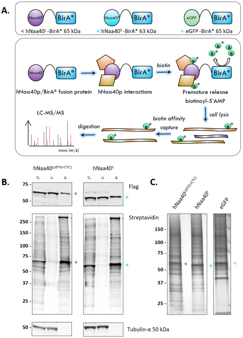
Figure 8.
Proximity-dependent biotin identification (BiolD) for the discovery of differential hNaa40p proteoform interactors in Flp-In T-REx 293 cells. (A) Overview of the BiolD (setups) and MS analysis workflow. Biological replicate samples were prepared for all 3 setups analyzed (i.e., biological replicate samples of stable cell lines with doxycycline (DOX) induced expression of eGFP, hNaa40L and hNaa40S BirA* fusion proteins) (B) Western blot analysis showing expression and (partial) biotinylation of hNaa40L and hNaa40S BirA* fusion proteins, and specific enrichment of biotinylated hNaa40p proteoforms following streptavidin affinity capture. TL = total lysate, U = unbound fraction and B = bound fraction. Purple, light blue and green arrows indicate hNaa40L, hNaa40S and eGFP-BirA* fusion proteins, respectively. The equivalents protein input of 50 and 200 µg ‘TL’ was analyzed for ‘TL’ and ’U’, and ‘B’ fractions, respectively. (C) Silver staining of biotinylated proteins following streptavidin purification (representative gel is shown). ~1.5% of total eluate sample corresponding to the input material of ~106 cells was analyzed.

