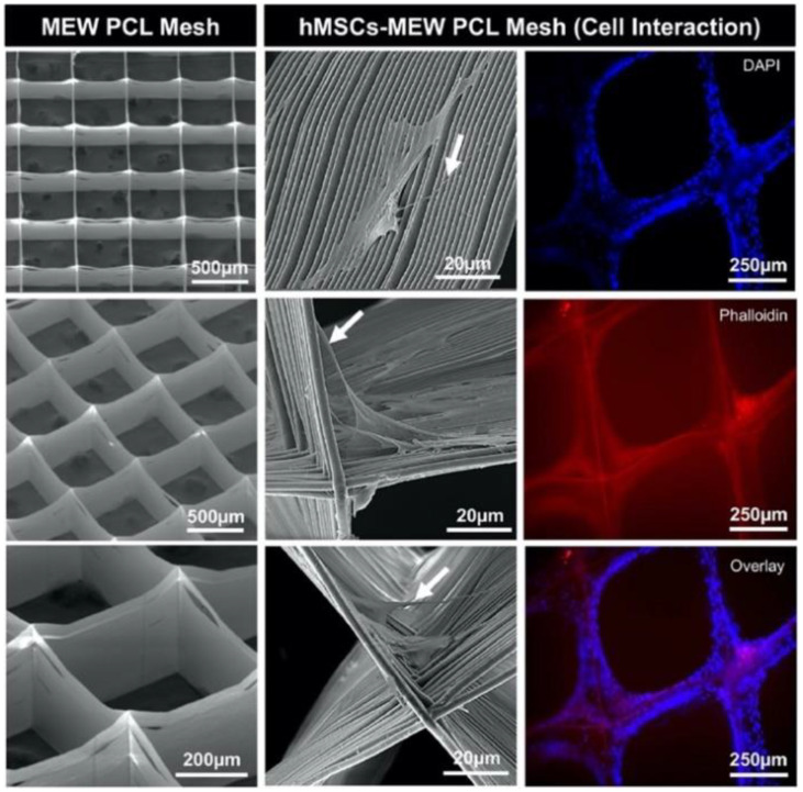
Figure 2.
(Left) Representative SEM micrographs of melt electrowriting (MEW) PCL mesh show the well-aligned (0–90°-oriented junctions) fibrous 3D architecture with a 500 μm pore size and a mean fiber diameter of 3.16 μm. (Middle) SEM micrographs of hMSCs-MEW PCL mesh interaction after 3 days of culture. Note significant cell attachment, proliferation, and protrusion along and around the printed PCL fibers. Filopodia are also indicated (white arrows). (Right) Fluorescence staining of hMSCs-MEW PCL mesh interaction after 3 days showing phalloidin (Red) staining of filamentous actin and DAPI (Blue) for the nucleus (for interpretation of the references to color in this figure legend, the reader is referred to the web version of this article). Reproduced with permission from Dubey, N et al., Acta Biomaterialia; published by Acta Materialia, Inc., 2020.

