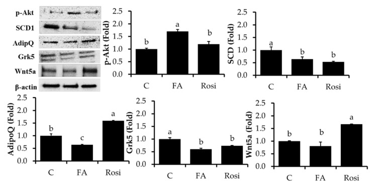
Figure 4.
Lipolysis-associated proteins were analyzed by immunoblotting. After incubation of 3T3-L1 adipocytes with/without FA and rosiglitazone (Rosi) at 10 μM treatment for 8 days, the expression levels of p-perilipin, p-HSL (Ser 565), p-AKT, SCD1, Wnt5a, adiponectin Grk5, and β-actin were measured. The results are represented as the mean ± standard error of the mean three replicates. The Fluorescence intensity of the targeted proteins was normalized with housekeeping protein (β-actin) using ImageJ software. a,b,c p < 0.05 statistically significant differences between control and treated groups.

