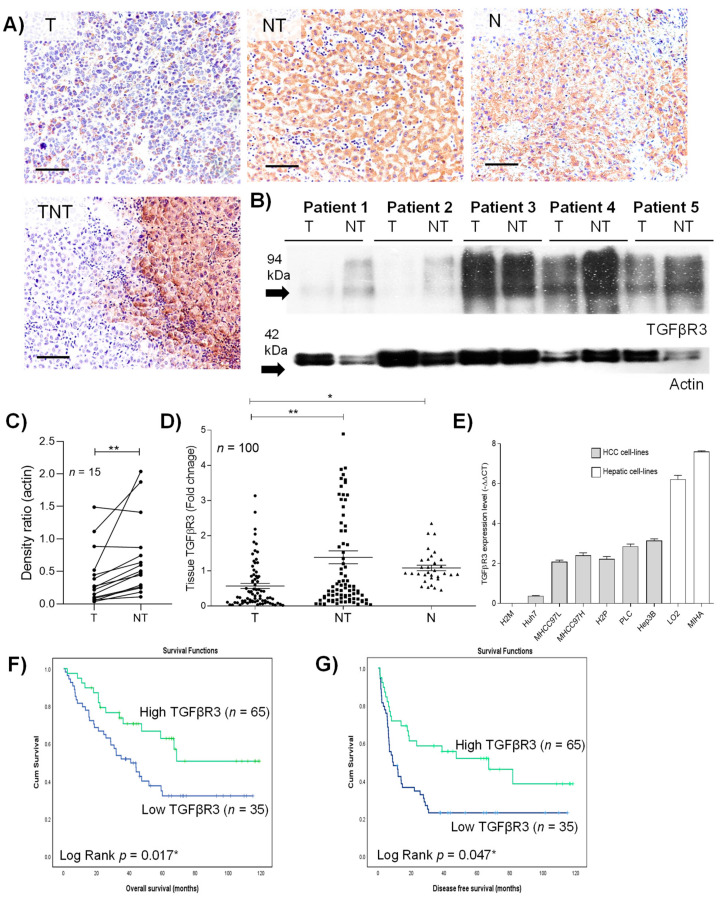
Figure 1.
Clinical characteristics of transforming growth factor beta type III receptor (TGFβR3) in hepatocellular carcinoma (HCC) tumoral tissue. (A) Representative image of immunohistochemical staining of TGFβR3. (B) Western blotting analysis of the protein level of TGFβR3. (C) Densitometric quantification of Western blotting relative to actin. (D) Quantitative-PCR analysis of transcript level of TGFβR3. (E) Transcript analysis of TGFβR3 in hepatocyte and HCC cell-lines. Kaplan–Meier analysis of (F) overall survival and (G) disease-free survival in HCC patients associated with the expression level of tumoral TGFβR3 transcript. T: tumor, NT: tumor-adjacent non-tumor, N: normal liver, TNT: intra- and peri-tumor; Scale bar: 50 μm; * p < 0.05, ** p < 0.01. Error bar indicated SEM. (Unpaired t-test for Figure 1C,D) (Paired t-test for Figure 1E) (Log rank test for Figure 1F,G).

