Figure 3.
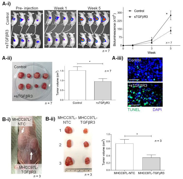
In vivo study of tumor-suppressive roles of (A) sTGFβR3 and (B) TGFβR3. (A) Male athymic mice bearing orthotopically grafted MHCC97L-Luciferase tumoral were injected with PBS (negative control) and 25 μg sTGFβR3 (n = 7) peritoneally weekly. (A-i) Monitoring of in situ tumor growth by Xenogen IVIS before and 1–5 week after injection with measurements of mean in vivo liver tumor bioluminescence of each group over time. Bioluminescent signals were quantified as photons/s at each imaging timepoint. (A-ii) Following euthanasia, tumor volume was examined and measured in each mouse. (A-iii) TUNEL staining of liver tumor tissues in the treatment and control group. (B-i) Subcutaneous tumor model setup of tumor nodule induced by MHCC97L-NTC (control) on the left flank and MHCC97L-TGFβR3 (knock-in) on the right flank in each mouse (n = 3). (B-ii) Tumor volume was examined and measured after scarification in week 4. * p < 0.05. Scale bar: 100 μm. Error bar indicated SEM. (Unpaired t-test).

