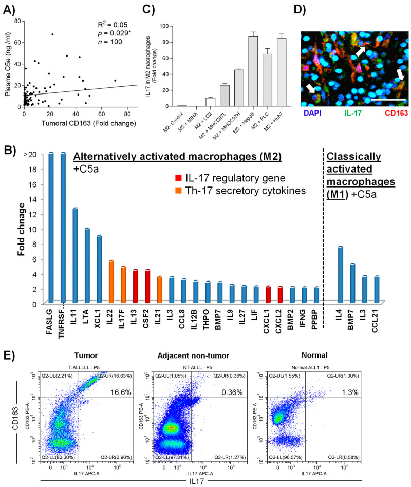
Figure 5.
The protumor functions of C5a via M2 macrophages activation. (A) Correlation between plasma C5a and scavenger receptor (SA) expression level in the tumoral tissues. (B) cDNA expression array analysis of M1 and M2 macrophages treated with recombinant C5a compared to untreated group. (C) Expression of IL-17 in M2 macrophages after incubation of HCC-conditioned medium. (D) Double immunofluorescence image of a clinical tumoral section labeled with antibodies against IL-17 (green) and CD163 (red). (E) Expression of surface CD163 and intra-cellular IL-17 in total CD45+ population of tumoral, adjacent non-tumoral tissue and healthy liver tissue isolated from HCC patients measured by flow cytometry. Scale bar: 100 μm. Error bar indicated SEM. (Chi-square test for Figure 5A)(Unpaired t-test for Figure 5C).

