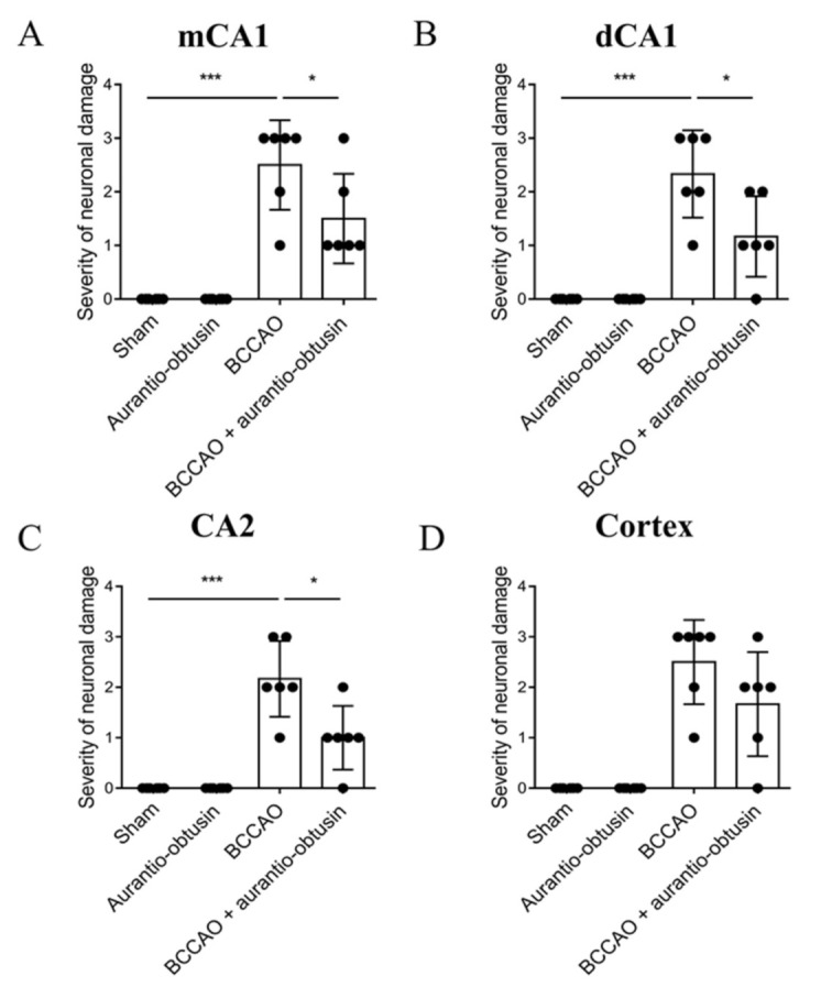
Figure 6.

Effect of aurantio-obtusin on transient forebrain ischemia-induced neuronal damage. (A−D). Severity of neuronal damage in mCA1 (A), dCA1 (B), CA2 (C), and cortex (D) regions. The cells were counted in six sections by every eight sections interval (total 48 sections) per animal by a person blind to the treatment group, and the average cell count per section was computed. The degree of damage by the Nissl staining after ischemia was semiquantitatively scored from 0 to 3. Neurons showing whole neuronal body shape were determined as healthy neurons. The percentage of healthy neurons compared to sham group was used as quantification criteria. (0, same to shame group; 1, >70% of sham group; 2, 40–70% of sham group; 3, 0–40% of sham group). Data represented as mean ± SD with raw data. * p < 0.05, *** p < 0.001.
