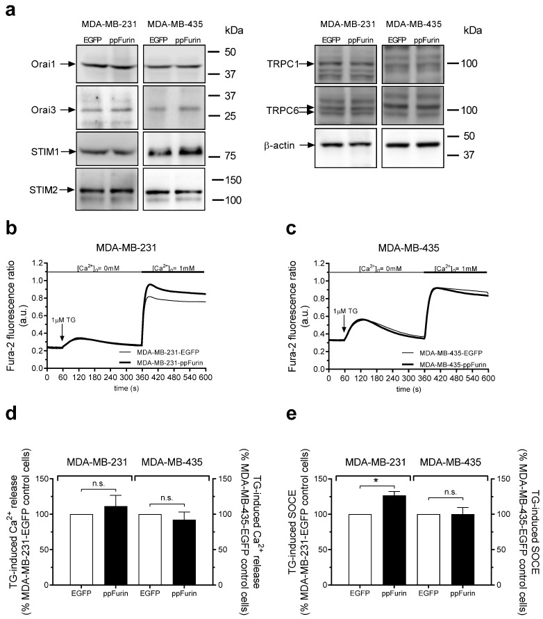
Figure 3.
ppFurin enhances store-operated Ca2+ entry (SOCE) in MDA-MB-231 cells. (a) Control MDA-MB-231 and MDA-MB-435 cells or expressing ppFurin were lysed and whole cell lysates were analyzed by Western blotting using anti-Orai1, Orai3, STIM1, STIM2, TRPC1, TRPC6, or β-actin antibody, as indicated. Blot images are from one experiment representative of three which gave similar results. (b–e). Changes in [Ca2+]I were detected in fura-2-loaded control MDA-MB-231 and MDA-MB-435 cells or expressing ppFurin. Cells were stimulated with 1 µM of TG in a Ca2+-free medium (100 µM of EGTA added), and later SOCE was detected by the addition of 1 mM of CaCl2 to the extracellular medium. (b,c) Traces are representative of 40 cells/day/3–5 days. (d,e) Histograms indicate TG-induced Ca2+ release and SOCE as the area under the curve, expressed as mean ± SEM and presented as percentage of their respective control cells. Statistical significance was assessed by Student’s t-test and * represents p < 0.05 as compared to cells transfected with pIRES2-EGFP empty vector.

