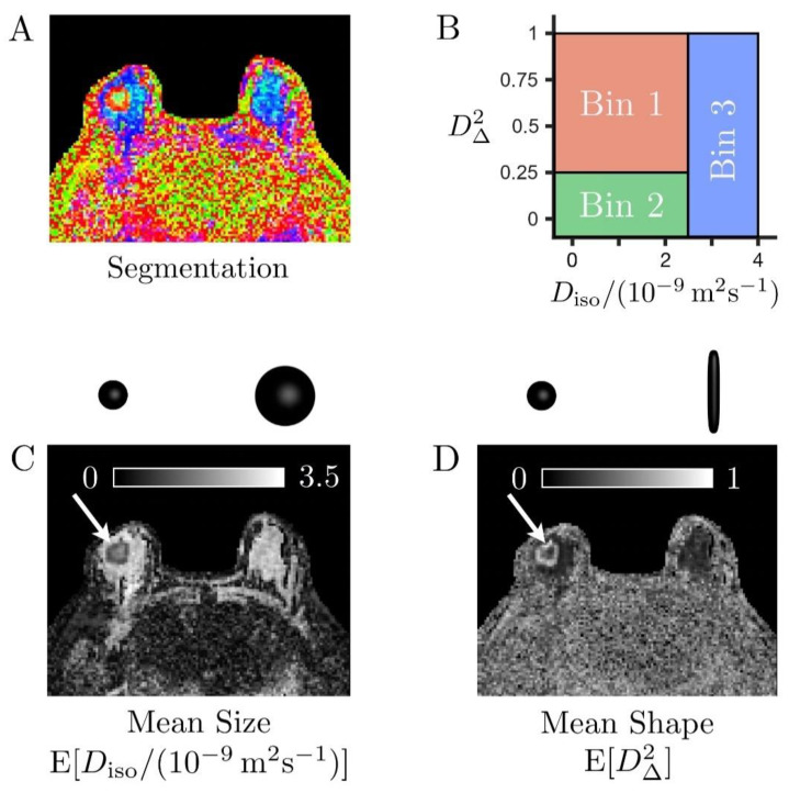
Figure 3.
Binning of the size–shape space of the diffusion tensor distributions (DTDs) in a case of invasive ductal carcinoma (A). Color-coded map derived from bin-resolved signal fractions, respectively associated with elongated cells (bin1, red), isotropic diffusion environments with low diffusivity (bin2, green), and isotropic diffusion environments with high diffusivity (bin3, blue) (B). Definition of the tissue-specific bins in the two-dimensional plane of tensor size Diso and squared tensor shape D∆2 (C,D). Grey scale maps reporting on the mean size E[Diso] and mean shape E[D∆2] of the entire voxel content, respectively. The cancer (white arrows) exhibits high cellularity (low mean size E[Diso]) compared to the healthy fibroglandular tissue (large blue and cyan areas indicated by the yellow arrow in (A). It also features prominent heterogeneity, with a core consisting of slow-diffusive isotropic environments (large fbin2, low mean shape E[D∆2]), which could correspond to densely packed isotropic cells or necrotic tissue, surrounded by a layer of elongated cells (large fbin1, high mean shape E[D∆2]).

