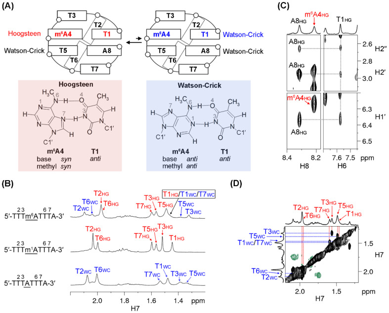
Figure 4.
(A) 5′-TTTm6ATTTA-3′ displayed a conformational exchange between two MDBs with T1·m6A4 Hoogsteen and Watson–Crick base pairs, respectively. (B) Comparison of the H7 signals of 5′-TTTm6ATTTA-3′ (top) with those in the m1A MDB (middle) and the TTTA MDB (bottom). The 1D 1H spectra were acquired at 0 °C. The subscripts “WC” and “HG” mean that T1·m6A4/ T1·m1A4/T1-A4 formed a Watson–Crick and Hoogsteen base pair respectively, in the corresponding structure. (C) NOEs of A8 H1′/H2′/H2′′-T1 H6 supported the formation of an MDB for the major conformer. The NOESY spectrum was acquired at 15 °C with a mixing time of 600 ms. (D) The H7 signals of the minor conformer were assigned based on their exchange cross peaks with the corresponding signals of the major conformer in a rotating frame Overhauser effect spectroscopy (ROESY) spectrum. The ROESY spectrum was acquired at 0 °C with a spinlock time of 200 ms.

