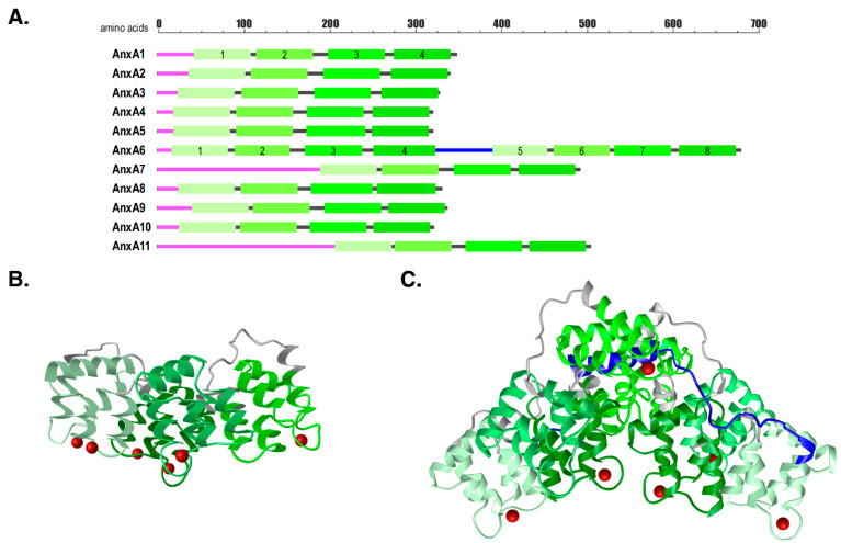
Figure 1.
Annexin structural organization. (A) Schematic overview of annexins. Magenta, N-terminal tails; light to dark green, C-terminal core domains with Annexin repeats 1–4 and 5–8 for AnxA6; grey, short spacer regions between tail and first Annexin repeat or between Annexin repeats; blue, AnxA6 linker region. (B) 3D-structures of human AnxA1 (PDB: 1AIN [17]) and (C) bovine AnxA6 (PDB: 1AVC [18]) cores (light to dark green, spacer regions in grey, AnxA6 linker region in blue), with coordinated calcium ions (red). 3D-structures are visualized with the iCn3D software vs. 2.24.6; (https://www.ncbi.nlm.nih.gov/Structure/icn3d/icn3d.html; accessed 12 March 2021).

