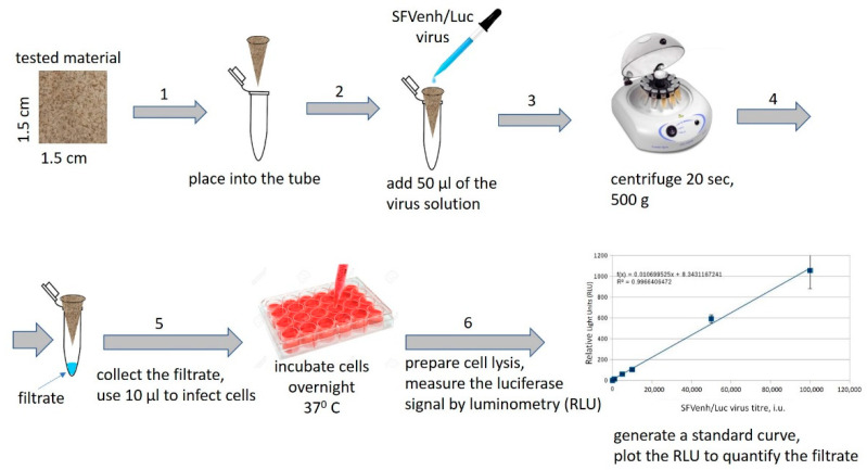
Figure 1.
Schematic representation of the virus filtration test (technical details are provided in text). (1) 1.5 cm × 1.5 cm filter sample is cut out from each material, folded into the form of a conical funnel, and placed into a 1.5 mL tube; (2) 50 µL of recombinant Semliki forest virus (SFV)-enh/Luc virus solution (107 i.u./mL) is added into the cone; (3) the tube is centrifuged to allow the virus to pass through the material; (4) the filtrate (indicated by arrow) is collected; (5) the filtrated is diluted and used for cell infection in a 24-well cell culture plate; (6) after overnight incubation of the plate the cell lysates are prepared and the virus infection is measured by detection of the luciferase activity in infected cells (luminometry). The cell infection with the standard dilutions of the virus is used to generate a standard curve and to calculate the amount of virus in the filtrate.

