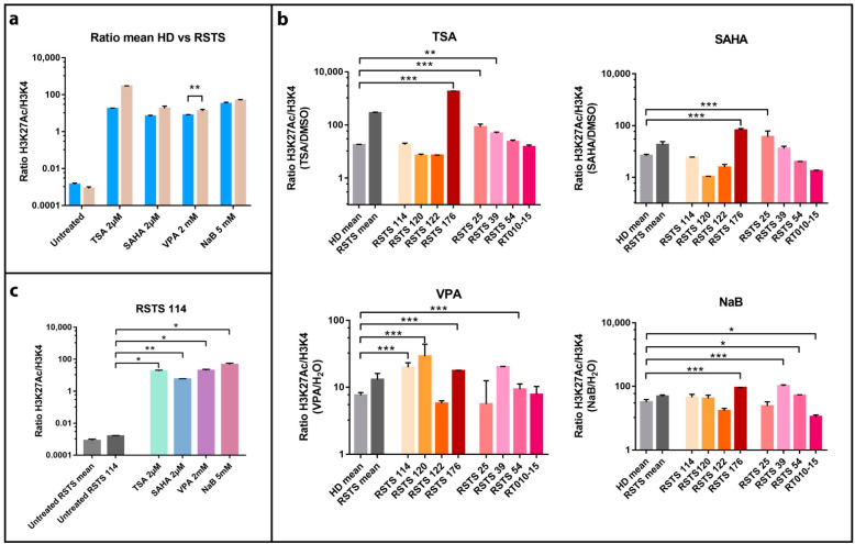
Figure 1.
Histone acetylation on Rubinstein–Taybi syndrome (RSTS) lymphoblastoid cell lines (LCLs) upon acetyltransferases (HAT) and deacetylases (HDAC) inhibitors exposure. H3K27 acetylation levels normalized on H3K4 unmodified, assessed by AlphaLISA®; levels of acetylation upon HDAC inhibitors (HDACi) are expressed as a ratio between the treatment and respective vehicle (HDACi/vehicle); on the Log scale, Y-axis H3K27 acetylation levels normalized, on X-axis lists of epigenetic treatments or untreated/treated single LCL or LCLs means. (a) Means of values of H3K27 acetylation in healthy donors (HD, in blue) and patients LCLs (RSTS, in pale brown) untreated and exposed to four different HDACi (trichostatin A (TSA) 2 µM, suberoylanilide hydroxamic acid (SAHA) 2 µM, valproic acid (VPA) 2 mM, and sodium butyrate (NaB) 5 mM). (b) H3K27 acetylation in eight RSTS LCLs (CREBBP LCLs in shades of red, EP300 LCLs in shades of pink) after exposure with the four different HDACi, compared to treated HD and RSTS means. (c) Insight on the single-patient response (RSTS 114) to the four compounds compared to untreated RSTS means and RSTS 114. Groups were compared using Student’s t-test as statistical method (* p < 0.05; ** p < 0.01; *** p < 0.001).

