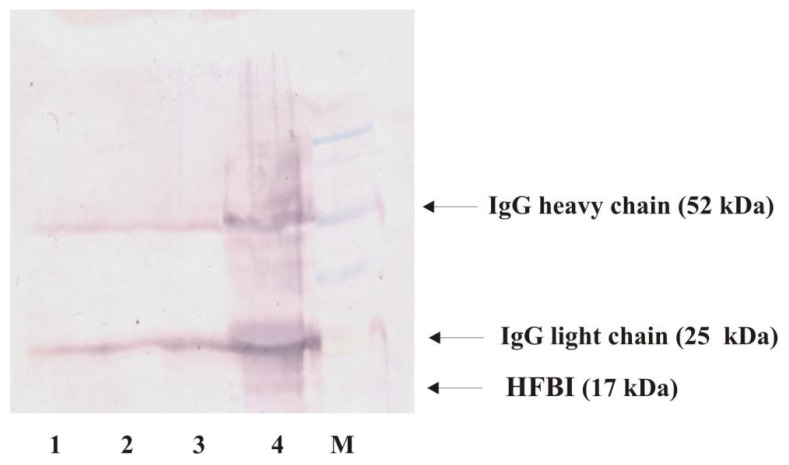
Figure 5.
Western blot analysis of hydrophobin. Proteins were immunoprecipitated using anti-HFBI antibody, and resulting pellets were washed and subjected to SDS-PAGE/Western blot analysis. Lane 1: liquid medium, lane 2: T. viride GZ1 culture, lane 3: liquid medium with PET, lane 4: T. viride GZ1 culture with PET, and M: molecular mass standard (BluEasy Prestained Protein Marker, Nippon Genetics, Dürren, Germany). All analyzed media/cultures were incubated for 3 months. Arrows indicate IgG heavy (52 kDa) and light (25 kDa) chains and protein that corresponds to HFBI (17 kDa).

