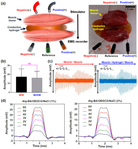
Figure 6.

Electrophysiological bridging effect of conductive hydrogel in ex vivo muscle defect model. (a) Schematic illustration (left) and photograph (right) of experimental setup for the EMG recording in two muscles spaced with the hydrogels. For EMG recording, three electrodes (anode, cathode, and reference) were utilized. (b) Average values of maximum EMG amplitude in muscle-to-muscle (M/M) or muscle-to-hydrogels-muscle (M/H/M) model during 8 V electrical stimulation (n = 3). (c) Pattern shapes of EMG signals in each M/M and the M/H/M model. (d) Comparison of the EMG amplitude monitored on the muscle spaced with the hydrogel prepared in NaCl 1% (left) or 2% (right) varying from 1 to 6 V electrical stimulation.
