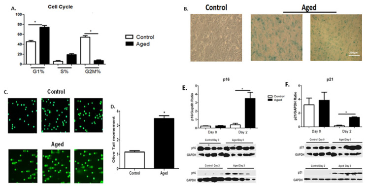
Figure 1.
(A) Effects of myoblast senescence on cell cycle progression. Percentages of cells in each phase arerepresented for the control and aged myoblasts. In contrast to the controls, the aged myoblasts exhibited an aging phenotype with an arrest of cell cycle progression, i.e., increased number of cells (%) in the G1 phase along with a reduction incell number in the G2/M phase. (B) Increased SA-β-gal activity as a result of myoblast aging. Representative images of the increased SA-β-gal-positive blue-green stain cells in the aged myoblast cultures. (C) Increased DNA damage as a result of myoblast aging. Representative alkaline comet assay images of aged myoblasts and control cells. (D) Quantification of the endogenous DNA damage in aged myoblasts compared to controls. (E,F) Effects of myoblast aging on the expression of cellular senescence-associated proteins p16 and p21. Three independent experiments were performed, and 200 cells per sample were scored. Representative Western blots and immunoblotting quantification of p16 (E) and p21 (F) expression in aged myoblasts compared to controls in the second day of their differentiation process. The expressions of the proteins were normalized to each corresponding GAPDH on the same immunoblot (Mean ± SE of 3 independent experiments performed in triplicate; * p < 0.05).

